PDF] Brain Tumor Segmentation of MRI Images Using Processed Image Driven U-Net Architecture
Por um escritor misterioso
Last updated 22 setembro 2024
![PDF] Brain Tumor Segmentation of MRI Images Using Processed Image Driven U-Net Architecture](https://d3i71xaburhd42.cloudfront.net/c750894747d2b3f841de55922b2b68794295de27/7-Table3-1.png)
A fully automatic methodology to handle the task of segmentation of gliomas in pre-operative MRI scans is developed using a U-Net-based deep learning model that reached high-performance accuracy on the BraTS 2018 training, validation, as well as testing dataset. Brain tumor segmentation seeks to separate healthy tissue from tumorous regions. This is an essential step in diagnosis and treatment planning to maximize the likelihood of successful treatment. Magnetic resonance imaging (MRI) provides detailed information about brain tumor anatomy, making it an important tool for effective diagnosis which is requisite to replace the existing manual detection system where patients rely on the skills and expertise of a human. In order to solve this problem, a brain tumor segmentation & detection system is proposed where experiments are tested on the collected BraTS 2018 dataset. This dataset contains four different MRI modalities for each patient as T1, T2, T1Gd, and FLAIR, and as an outcome, a segmented image and ground truth of tumor segmentation, i.e., class label, is provided. A fully automatic methodology to handle the task of segmentation of gliomas in pre-operative MRI scans is developed using a U-Net-based deep learning model. The first step is to transform input image data, which is further processed through various techniques—subset division, narrow object region, category brain slicing, watershed algorithm, and feature scaling was done. All these steps are implied before entering data into the U-Net Deep learning model. The U-Net Deep learning model is used to perform pixel label segmentation on the segment tumor region. The algorithm reached high-performance accuracy on the BraTS 2018 training, validation, as well as testing dataset. The proposed model achieved a dice coefficient of 0.9815, 0.9844, 0.9804, and 0.9954 on the testing dataset for sets HGG-1, HGG-2, HGG-3, and LGG-1, respectively.
![PDF] Brain Tumor Segmentation of MRI Images Using Processed Image Driven U-Net Architecture](https://www.science.org/cms/10.1126/sciadv.add3607/asset/f006810b-5ff3-4034-a144-00b49132fbcb/assets/images/large/sciadv.add3607-f1.jpg)
SynthSR: A public AI tool to turn heterogeneous clinical brain scans into high-resolution T1-weighted images for 3D morphometry
![PDF] Brain Tumor Segmentation of MRI Images Using Processed Image Driven U-Net Architecture](https://media.springernature.com/m685/springer-static/image/art%3A10.1038%2Fs41598-023-47107-7/MediaObjects/41598_2023_47107_Fig1_HTML.png)
Utilizing deep learning via the 3D U-net neural network for the delineation of brain stroke lesions in MRI image
![PDF] Brain Tumor Segmentation of MRI Images Using Processed Image Driven U-Net Architecture](https://image.isu.pub/221203102411-2d295fd32a8526195049b137eb92dbb6/jpg/page_1_thumb_large.jpg)
Unet 3+ For Brain Tumor Segmentation : A Study by IRJET Journal - Issuu
![PDF] Brain Tumor Segmentation of MRI Images Using Processed Image Driven U-Net Architecture](https://media.springernature.com/m685/springer-static/image/art%3A10.1186%2Fs12859-021-04347-6/MediaObjects/12859_2021_4347_Fig4_HTML.png)
MRI-based brain tumor segmentation using FPGA-accelerated neural network, BMC Bioinformatics
![PDF] Brain Tumor Segmentation of MRI Images Using Processed Image Driven U-Net Architecture](https://miro.medium.com/v2/resize:fit:1358/1*Mbdtdnni6jVaPp5JCuJOVg.png)
Brain Tumor Segmentation with U-Net in Python: A Deep Learning Approach, by Lyron Foster
![PDF] Brain Tumor Segmentation of MRI Images Using Processed Image Driven U-Net Architecture](https://media.springernature.com/m685/springer-static/image/art%3A10.1186%2Fs12859-021-04347-6/MediaObjects/12859_2021_4347_Fig1_HTML.png)
MRI-based brain tumor segmentation using FPGA-accelerated neural network, BMC Bioinformatics
![PDF] Brain Tumor Segmentation of MRI Images Using Processed Image Driven U-Net Architecture](https://www.mdpi.com/electronics/electronics-09-02203/article_deploy/html/images/electronics-09-02203-g001.png)
Electronics, Free Full-Text
![PDF] Brain Tumor Segmentation of MRI Images Using Processed Image Driven U-Net Architecture](https://media.arxiv-vanity.com/render-output/7558552/graph1.jpg)
BiTr-Unet: a CNN-Transformer Combined Network for MRI Brain Tumor Segmentation – arXiv Vanity
![PDF] Brain Tumor Segmentation of MRI Images Using Processed Image Driven U-Net Architecture](https://production-media.paperswithcode.com/tasks/brain-tumor-segmentation_Omf9jBU.png)
Brain Tumor Segmentation
![PDF] Brain Tumor Segmentation of MRI Images Using Processed Image Driven U-Net Architecture](https://www.degruyter.com/document/doi/10.1515/jisys-2022-0206/asset/graphic/j_jisys-2022-0206_fig_006.jpg)
A novel deep learning-based brain tumor detection using the Bagging ensemble with K-nearest neighbor
Recomendado para você
-
 Brain Test Level 191 I hate this! The baby is crying again! Stop this scream in 202322 setembro 2024
 -
 Brain Test Level 191 Walkthrough Solution22 setembro 2024
 -
 Brain Test ○ Nível 19122 setembro 2024
 -
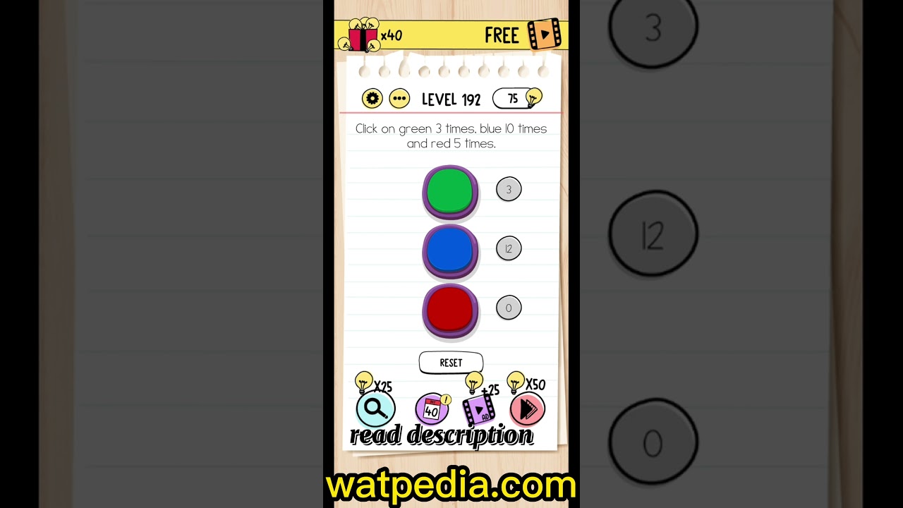 Brain Test Level 192 Click on green 3 times, blue 10 times and red 5 times ✓22 setembro 2024
 -
 Thank you u/Bogi_D for signing my #1. I'm a cage person now! : r/CollectibleAvatars22 setembro 2024
 -
 Mind-Mapping — Cajun Koi Academy22 setembro 2024
 -
 Astronomy UV beads Solar Beads Lab Help Stop Skin Cancer Middle School Science - Classful22 setembro 2024
 -
 1.6: Communicating Scientific Discoveries to Peers - Social Sci LibreTexts22 setembro 2024
 -
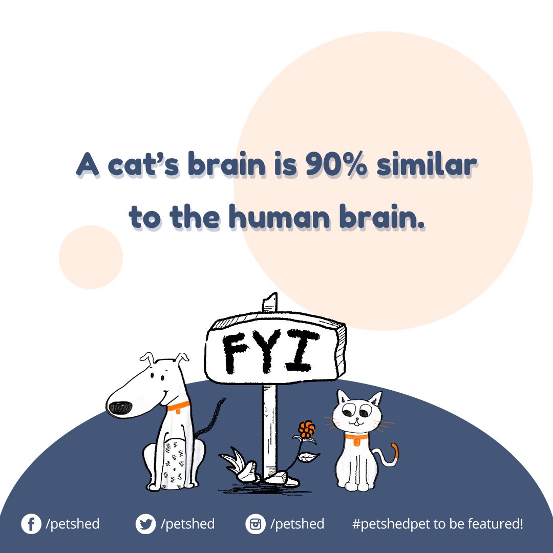 Pet Shed (@PetShed) / X22 setembro 2024
 -
 Brain Test 4: Tricky Friends Level 191-200 Walkthrough Solution (NEW UPDATE)22 setembro 2024
você pode gostar
-
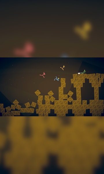
Buy Stick Fight: The Game Steam Key NORTH AMERICA - Cheap - !22 setembro 2024
-
 Car Park at Helmont House - from £1.50/hour22 setembro 2024
 -
 Baki Hanma Anime Confirms Returning Lead Actor, Posts Art by22 setembro 2024
 -
 Millwall 2-0 Middlesbrough: Zian Flemming double steers Lions to victory, Football News22 setembro 2024
 -
 Wake Up Aid Bed Ladder Moving Assist Belt Hoist Gait Belt Walker Framing Tools Twin Floor Bed Frame Pull Up Straps Kit Bed Pull Up Belt Bed Support Belt Black Elderly22 setembro 2024
 -
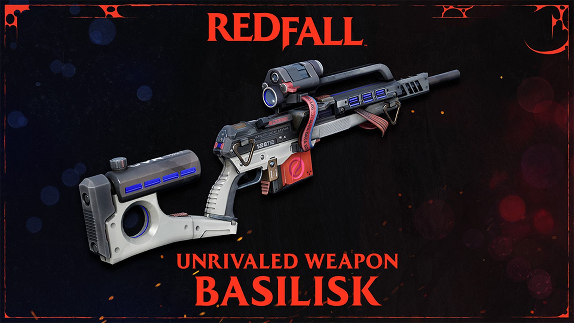 Redfall Title Update 3 is Live, Adds Unrivaled Sniper Rifle “Basilisk”22 setembro 2024
 -
![Building a multiplayer tic-tac-toe game - Socket.IO Cookbook [Book]](https://www.oreilly.com/api/v2/epubs/9781785880865/files/graphics/B04893_03_03.jpg) Building a multiplayer tic-tac-toe game - Socket.IO Cookbook [Book]22 setembro 2024
Building a multiplayer tic-tac-toe game - Socket.IO Cookbook [Book]22 setembro 2024 -
 UCN Fredbear : r/fivenightsatfreddys22 setembro 2024
 -
 Meikyuu Black Company Review22 setembro 2024
 -
 Dig Dig by elysiaisalive22 setembro 2024